Call us now :07971671031

 Send Inquiry
Trinocular Research Microscope
Trinocular Research Microscope Specification
- Features
- Heavy metallic body, anti-fungal optical coating, coaxial stage movement
- Spare Parts
- Power cord, fuse, dust cover, immersion oil, lamp, spare bulb
- Focus System
- Coaxial coarse & fine adjustment knobs on both sides
- View Head
- Trinocular, inclined at 45, rotatable 360
- Theory
- Compound Microscope for research applications
- Drawtube
- Trinocular
- Sensor
- Not Applicable (optical microscopy)
- Resolution
- Optical; depends on objective, up to 0.2 m
- Interface
- Tri-port for camera/eyepiece/observation
- Frame Rate
- Dependent on camera used (not applicable for optical only)
- Magnification
- 40x to 1000x (using various objectives and eyepieces)
- Eyepieces
- Wide Field 10x paired
- Eyepiece Tube
- Inclined at 45, 360 rotatable
- Illumination
- Built-in LED or Halogen 6V/20W illuminator (with intensity control)
- Coarse Adjustment Range
- 15 mm
- Fine Adjustment Range
- 0.002 mm precision
- Still Image Capture Resolution
- Depends on camera; not included as standard
- Video Capture Resolution
- Depends on camera; not included as standard
- Image Format
- Direct optical view; camera required for digital capture
- Objective Achromatic
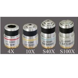
- Achromatic Objectives 4x, 10x, 40x (spring), 100x (oil, spring) standard
- Condenser
- Abbe condenser N.A. 1.25 with iris diaphragm and filter holder
- Light Source
- Halogen Lamp 6V/20W or LED as per model
- Head Rotation
- 360 rotatable for convenient sharing and demonstration
- Voltage
- 220V AC/50 Hz standard
- Safety
- Fitted with voltage protector and thermal fuse
- Filter Holder
- Provided with blue, green, and ground glass filters
- Stage Movement
- Racks and pinion, low-position coaxial controls
- Body Construction
- Robust anti-vibration metallic base designed for stability
- Color
- White and black finish (as per image provided)
- Nosepiece
- Quadruple reverse ball bearing type for smooth objective change
- Diopter Adjustment
- On left eyepiece to compensate for individual vision variation
About Trinocular Research Microscope
With the excellent support of our experienced workforce, we have been indulged in manufacturing, exporting and supplying our clients a comprehensive array of Trinocular Research Microscope. This microscope is manufactured with the aid of the quality approved components by following the predefined industry terms. Ideal for clinical laboratories, college and school laboratories, offered microscope is used to view cell structures, tissue culture samples and living microorganisms. Moreover, this Trinocular Research Microscope is available in different specifications at nominal rates.
Features:
- Flexible light source
- Ergonomic design
- Maintenance free
- Compact and light in weight
Specifications
|
Objective/Eyepiece |
5X |
10X |
45X |
100X |
|
10X W.F |
15X |
100X |
450X |
1000X |
|
15X HUY.(Optional) |
75X |
150X |
675X |
1500X |
|
Accessories
- Vinyl cover
- Cleaning Brush
- Duster
Advanced Optical Performance
Equipped with achromatic objectives (4x, 10x, 40x spring, 100x oil spring) and an Abbe condenser (N.A. 1.25), this microscope guarantees clear, sharp imaging suited for detailed research. With a wide magnification range and anti-fungal coatings, users can maintain precision over extended use.
User-Friendly Ergonomics
Designed with a 360 rotatable viewing head and 45 inclined eyepiece tubes, the microscope accommodates multiple users and minimizes fatigue. The mechanical stage's low-position controls offer efficient, comfortable operation for lengthy observation sessions.
Robust Construction & Safety
A heavy metallic body and anti-vibration base provide durability and stability. Integrated safety measures-such as a voltage protector and thermal fuse-ensure reliable operation, while essential spares like fuses and bulbs support maintenance.
FAQ's of Trinocular Research Microscope:
Q: How does the anti-vibration base contribute to reliable observations?
A: The robust metallic base minimizes vibrations, reducing image distortion and stabilizing your observations, especially at high magnifications. This is vital for consistent results in precise research work.Q: What is the benefit of the trinocular head and how can it be utilized?
A: The trinocular head allows for simultaneous observation through the eyepieces and camera port, facilitating digital imaging, documentation, and live demonstration. Its 360 rotation is ideal for classroom sharing or collaborative lab work.Q: When should you use the diopter adjustment on the left eyepiece?
A: Diopter adjustment compensates for differences in vision between your eyes. Use this feature whenever you or another user feels images are out of focus, even after standard focussing, to achieve the clearest view for individual vision.Q: Where can the filters be installed, and what is their purpose?
A: The provided blue, green, and ground glass filters are placed in the dedicated filter holder under the condenser. They enhance image contrast, improve color rendering, and protect the eyes from intense illumination.Q: What is the process for changing objectives with the quadruple nosepiece?
A: Simply rotate the reverse ball bearing nosepiece to smoothly switch between the four achromatic objectives. The ball bearing mechanism ensures accurate alignment and ease of use, even during frequent changes.Q: How does the illumination system improve observation quality?
A: The built-in LED or halogen lamp offers consistent, adjustable lighting, ensuring that your specimens are brightly and evenly illuminated. This is crucial for observing fine details under various magnifications.Q: What are the main advantages of using this microscope for research applications?
A: Its heavy-duty build, advanced optical system, ergonomic design, and integrated safety features make it ideal for professional labs. The trinocular interface supports both direct viewing and high-quality digital documentation, enhancing both research productivity and collaborative study.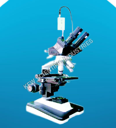
Tell us about your requirement

Price:
Quantity
Select Unit
- 50
- 100
- 200
- 250
- 500
- 1000+
Additional detail
Mobile number
Email

More Products in Microscopes Category
Objective Microscope
Drawtube : Other, Standard monocular drawtube compatible with most laboratory setups.
Illumination : Halogen illuminator, 20W lamp for uniform lighting.
Eyepieces : Widefield 10X eyepiece supplied.
Focus System : Coarse and fine adjustment knobs; vertical movement of stage.
View Head : Monocular, inclined 45
Light Source : Builtin halogen lamp, 20W, adjustable brightness.
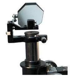
Camera Lucida (Mirror Type)
Drawtube : Other, Compatible with standard 23mm or 30mm inner diameter microscope drawtubes.
Illumination : External light source recommended; device itself does not provide illumination.
Eyepieces : Fits standard microscope eyepiece sizes (typically 10x or 15x).
Focus System : Manual via microscope focusing system.
View Head : Single viewing arrangement through optical system.
Light Source : Ambient or external illumination.
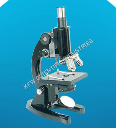
Student Microscope
Drawtube : Monocular
Illumination : Planoconcave mirror, substage illumination (natural or artificial)
Eyepieces : Wide field 10x
Focus System : Coarse and fine focusing knobs
View Head : Monocular, 45 inclined
Light Source : Plane & Concave Mirror for light reflection
Eyepieces Huygenian Microscope
Drawtube : Other, Standard mechanical drawtube compatible with compound microscopes.
Illumination : Depends on microscope; eyepiece does not provide illumination.
Eyepieces : Huygenian type, doublelens construction; available in various magnifications.
Focus System : Manual focus via microscope system.
View Head : Suitable for monocular, binocular, and trinocular heads depending on microscope compatibility.
Light Source : Eyepiece does not have its own light source; illumination is via microscope lamp or mirror.
Our Products
- Slit Lamp Ophthalmic Equipment
- Slit Lamp
- Surgical Equipments
- Laboratories Instruments Equipment
- Microscopes
- Scientific Instruments
- ENT Equipment
- Pharmacy instrument equipment
- Pathological Equipment
- Physics Lab Equipments
- Physics instruments
- Laboratory Glassware
- Digital Polarimeter
- Microprocessor Viscometer
- Double Beam UV-VIS Spectrophotometer
- Single Beam UV-Vis Spectrophotometer
- Kitchen equipments
- Dental Equipment
- Chapati Making Machines
- Water Heating System OR Hamam
- High Performance Liquid Chromatography Systems
- Chemistry
- Equipment Accessories
- Gynecology Equipment


B.O : 5430/3, Anaj Mandi, Science Market, Near Nigar Cinema,
R.O : 102-B Ram Nagar,Ambala Cantt - 133001, Haryana, India

Mr VISHAL KUMAR
(Managing Director)
Mobile :07971671031
 Send Inquiry
Developed and Managed by Infocom Network Private Limited.
